Muscular control of the eyes
Six muscles attached to the outer surface of each eye
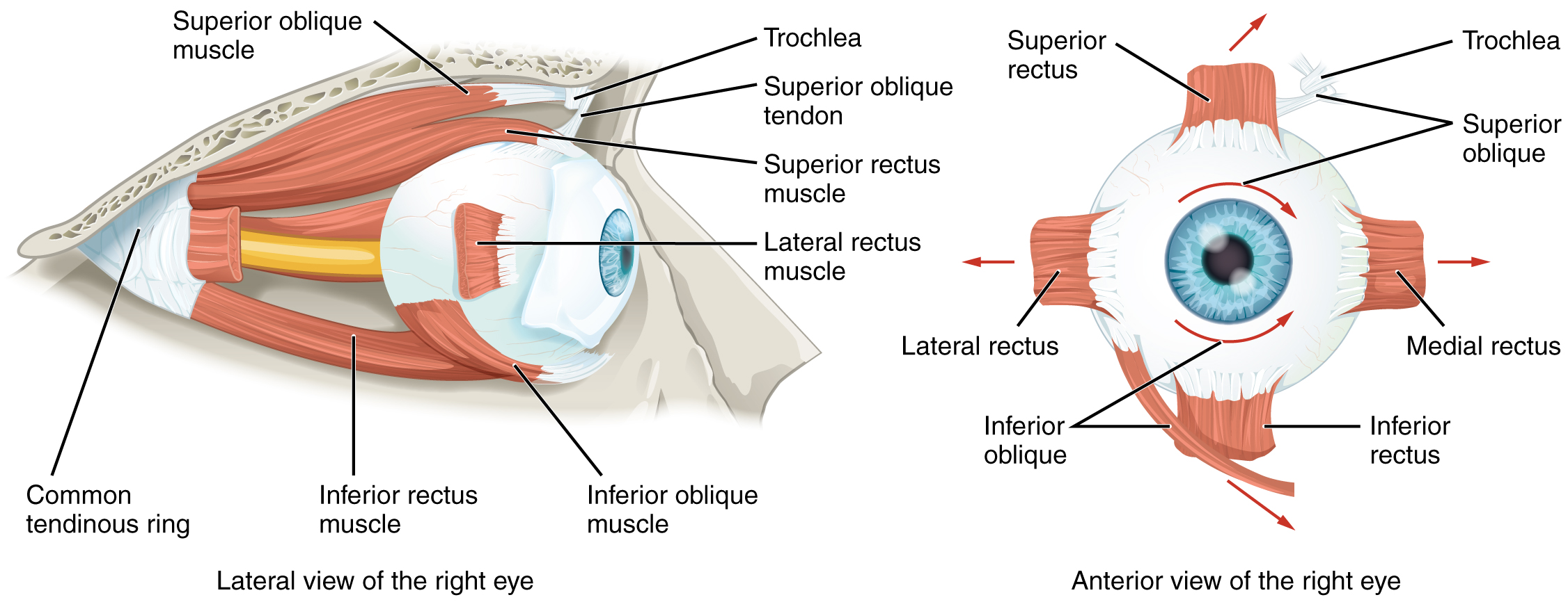
Each muscle pair is mostly associated with one movment type:
However, in practice, all muscles contribute to all movements.
The eye as a dipole
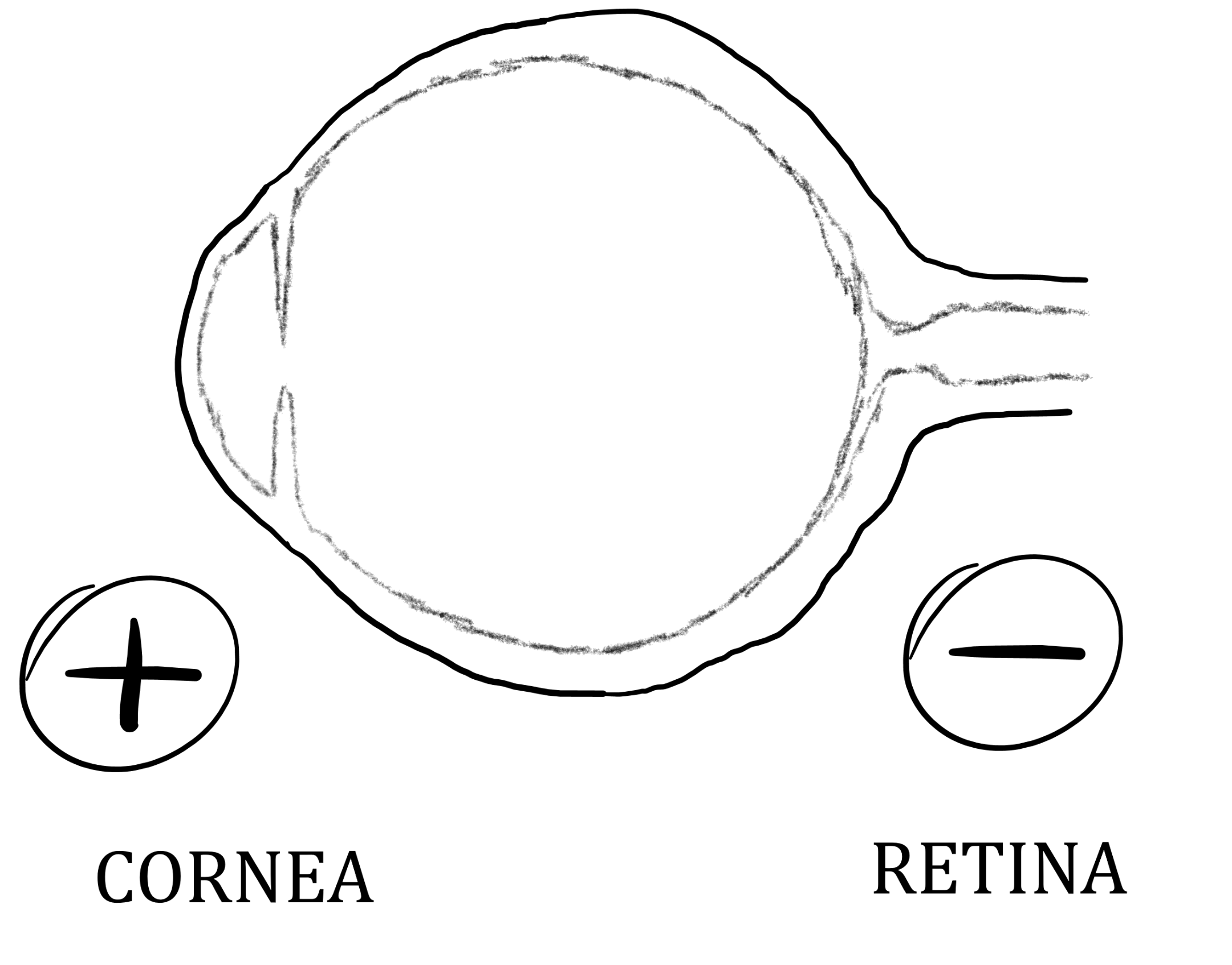
The retina (or fundus) is electrically more negatively charged than the cornea.
Why? The greater negativity in the retina is due to metabolic activity due to photoreceptors and other neurons.
This creates a potential, called corneoretinal (or corneofundal) potential. This potential forms a dipole.
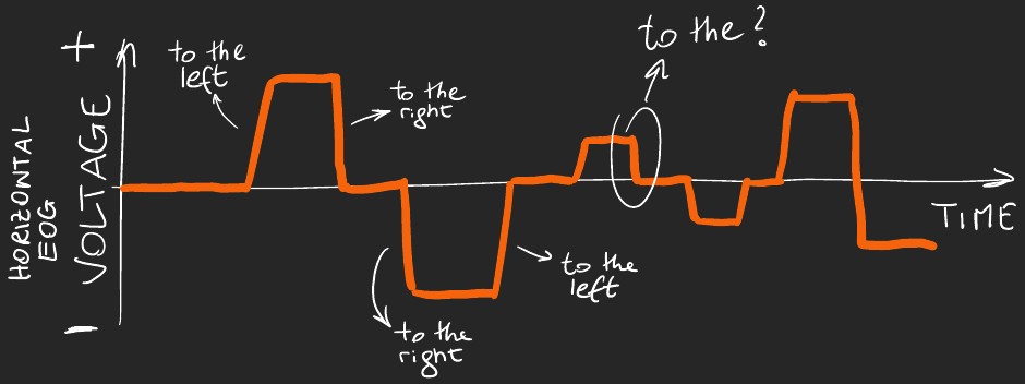
Measuring changes to the dipole
Assume you place two electrodes, one left and one right of the eyes.
When you look left: the left side becomes more positive while the right side becomes more negative.

When you look right: the right side becomes more positive while the left side becomes more negative.

Electrooculography (EOG)
Electro = electrical activity; oculo = eyes; graphy = measurement
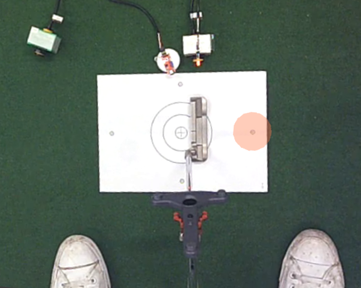
A system consisting of electrodes (plus electrolytic paste, other hardware, and software–more on this later) to record electrical activity near the eyes.

Electrooculography (EOG)
Electro = electrical activity; oculo = eyes; graphy = measurement
A system consisting of electrodes (plus electrolytic paste, other hardware, and software–more on this later) to record electrical activity near the eyes.

Electrooculography (EOG)
Electro = electrical activity; oculo = eyes; graphy = measurement
A system consisting of electrodes (plus electrolytic paste, other hardware, and software–more on this later) to record electrical activity near the eyes.

Electrooculography (EOG)
Electro = electrical activity; oculo = eyes; graphy = measurement
A system consisting of electrodes (plus electrolytic paste, other hardware, and software–more on this later) to record electrical activity near the eyes.

Electrooculography (EOG)
Electro = electrical activity; oculo = eyes; graphy = measurement
A system consisting of electrodes (plus electrolytic paste, other hardware, and software–more on this later) to record electrical activity near the eyes.

Electrooculographic signals #1
Electrode locations
Electrodes are placed around the eyes, but more specifically where?
It depends on the direction in which we want to record eye movements.

Electrode locations
Electrodes are placed around the eyes, but more specifically where?
It depends on the direction in which we want to record eye movements.

Electrode locations
Electrodes are placed around the eyes, but more specifically where?
It depends on the direction in which we want to record eye movements.

Electrode location nomenclature
There is a standardized nomenclature to identify the location of the electrodes.
- “L” stands for lateral
- “I” stands for inferior (below)
- “S” stands for superior (above)
- “1” stands for left
- “2” stands for right
| LABEL | SITE |
|---|---|
| LO1 | lateral ocular left |
| LO2 | lateral ocular right |
| IO1 | inferior ocular left |
| IO2 | inferior ocular right |
| SO1 | superior ocular left |
| SO2 | superior ocular right |

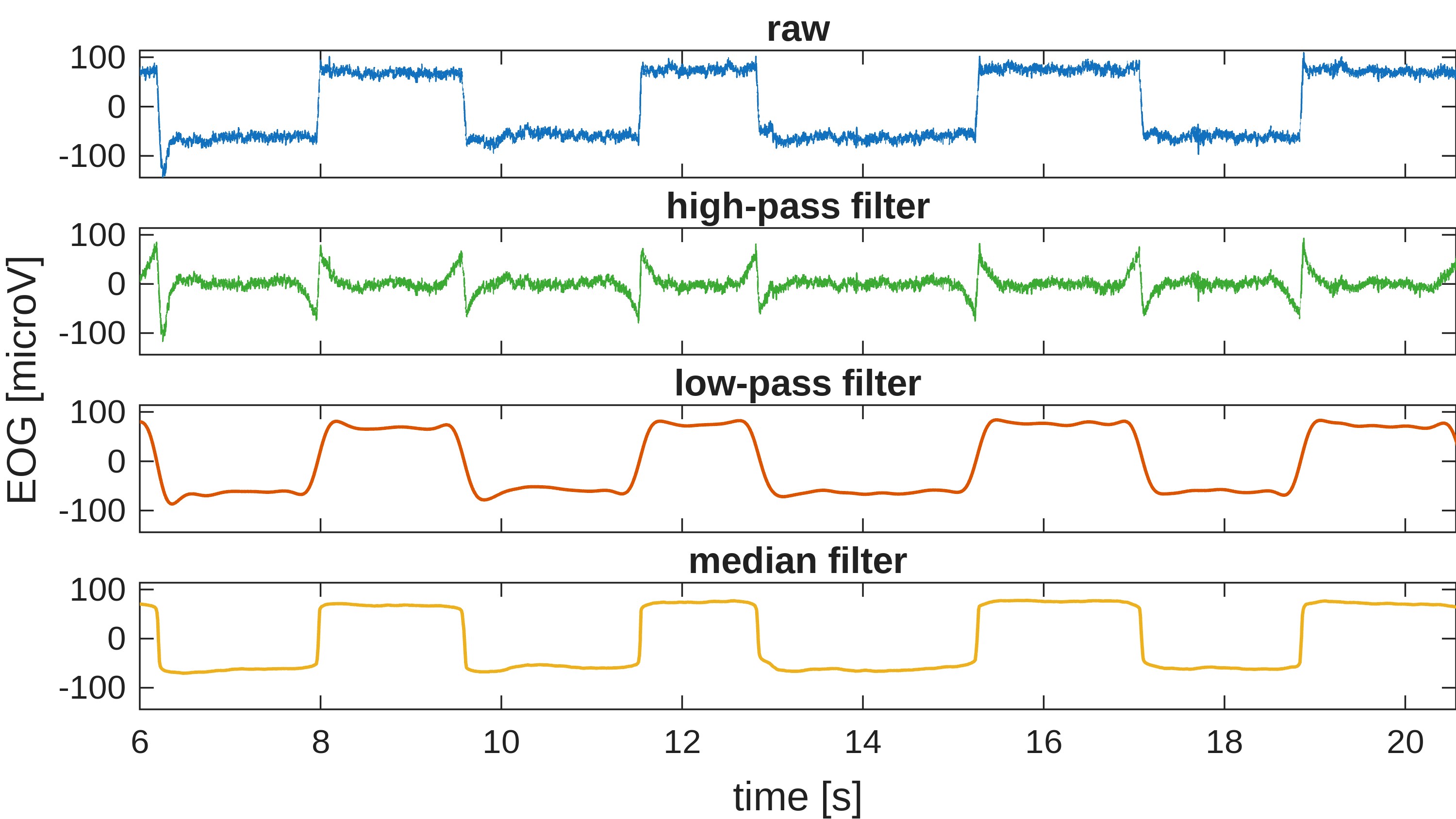
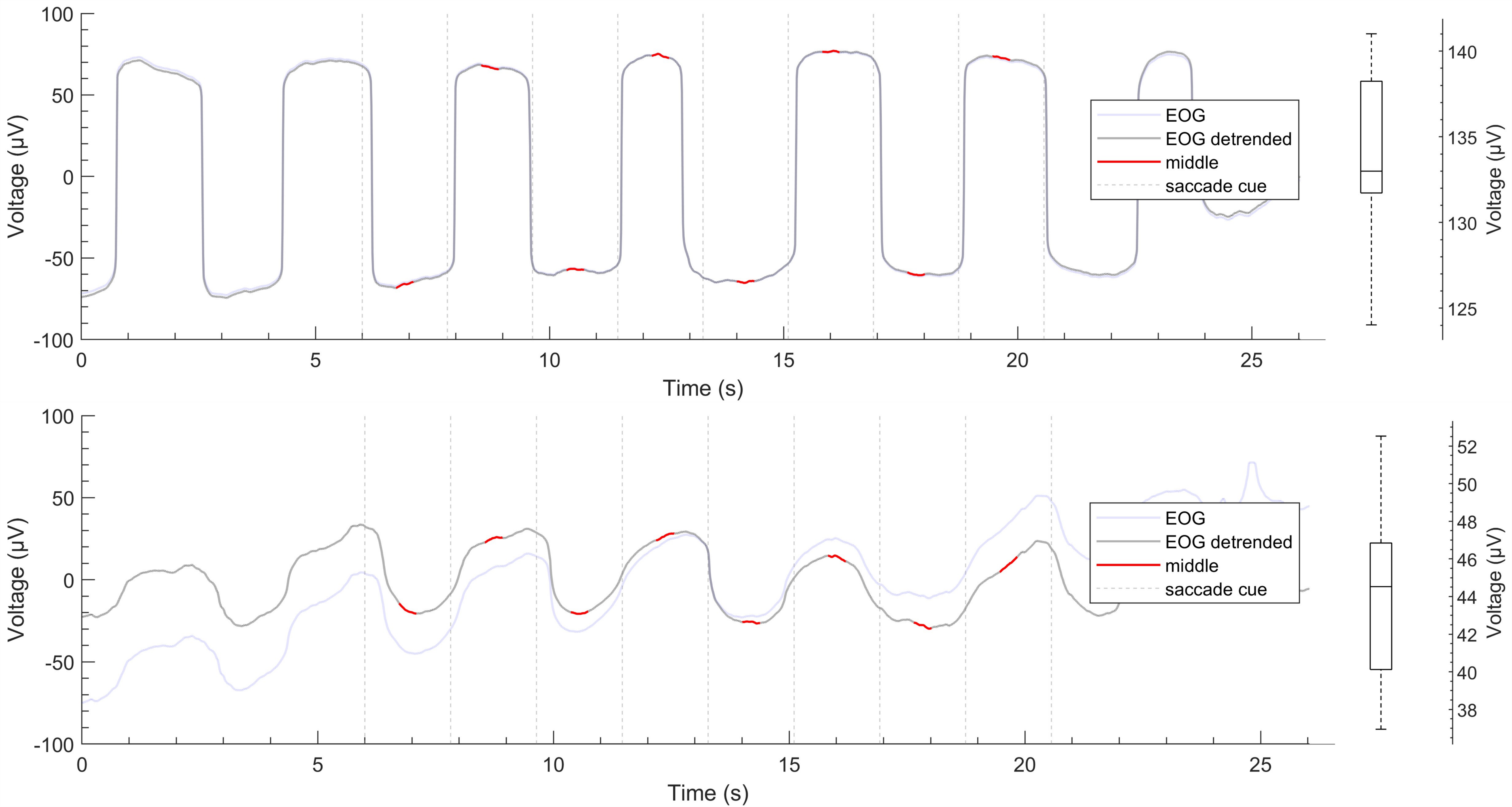
Impact of high-pass filtering
High-pass filtering is not recommended when studying eye movements. But many systems apply high-pass filtering (it’s simpler).
The high-pass filter reduces slow trends and retains faster changes.
Consider an EOG signal (with saccades and fixations). Applying high-pass filter makes the EOG signal less “square”. This is as if only the initial changes are detected or only the quick changes that happen (keeping eyes still can be considered like an extremely slow change).
For example, voltage increases as you move the eyes up. You would expect the voltage to not change is you maintain your eyes in that position. But when high-pass filtering is applied, the voltage goes gradually back to baseline even if the eyes are still up.
EOG calibration
The native units of the EOG are Volts, but eye movements are better described as visual angles. Eye movements are angular in nature.
Calibration allows to calculate how many Volts correspond to 1 degree of visual angle, so that we can convert our EOG recording from Volts to degrees.
Calibrated EOG
The EOG signal is now represented in degrees of visual angle.
The figure shows horizontal, vertical, and combined EOG signals, in the seconds before the execution of a golf putt.
The combined signal is computed as the vectorial sum of the calibrated horizontal and vertical signals.
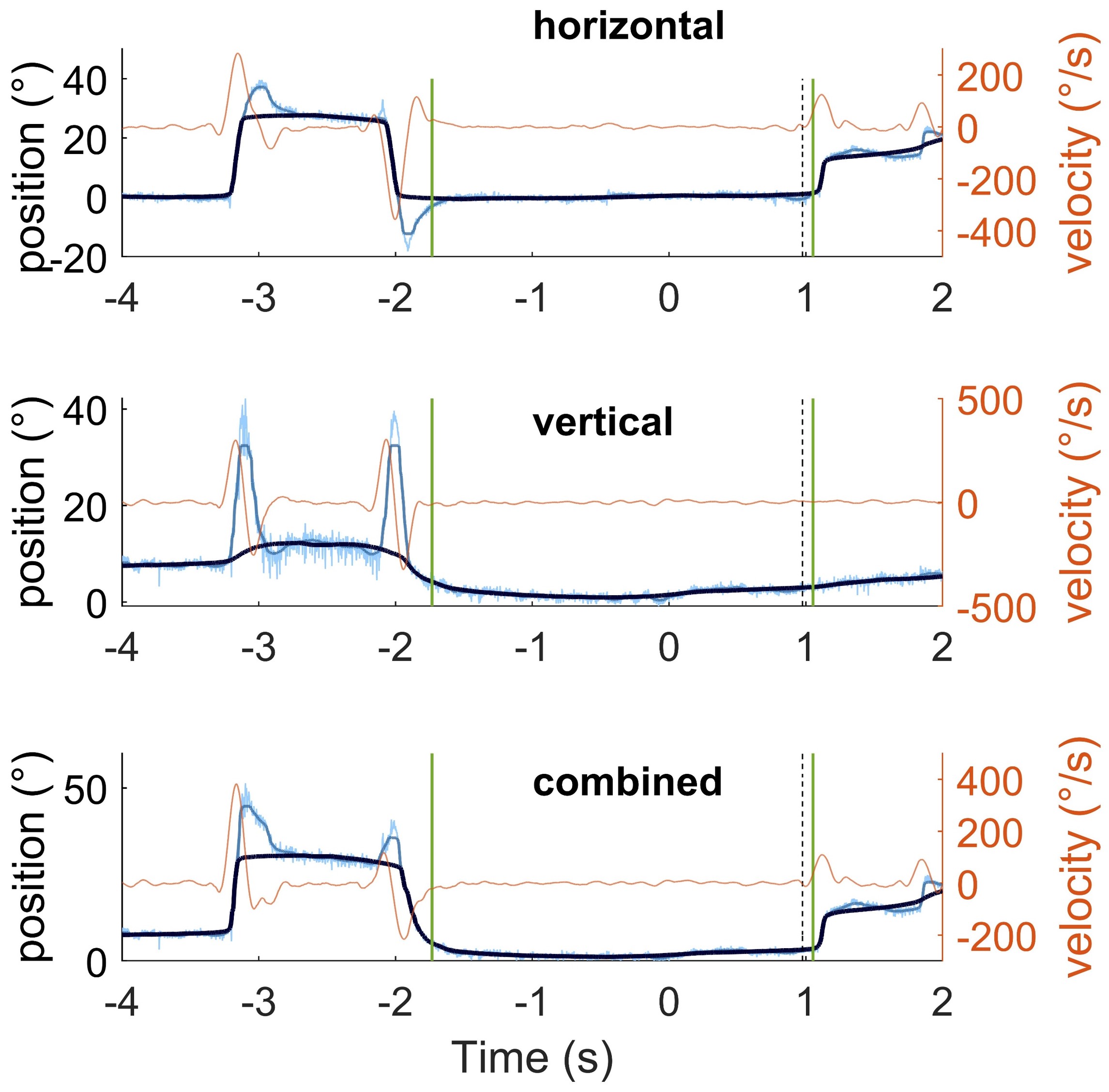
Because the source of the EOG (the corneoretinal potential) is not fixed, calibration should be done regularly unless the recording session is very short.
Eye blink as seen in the EOG
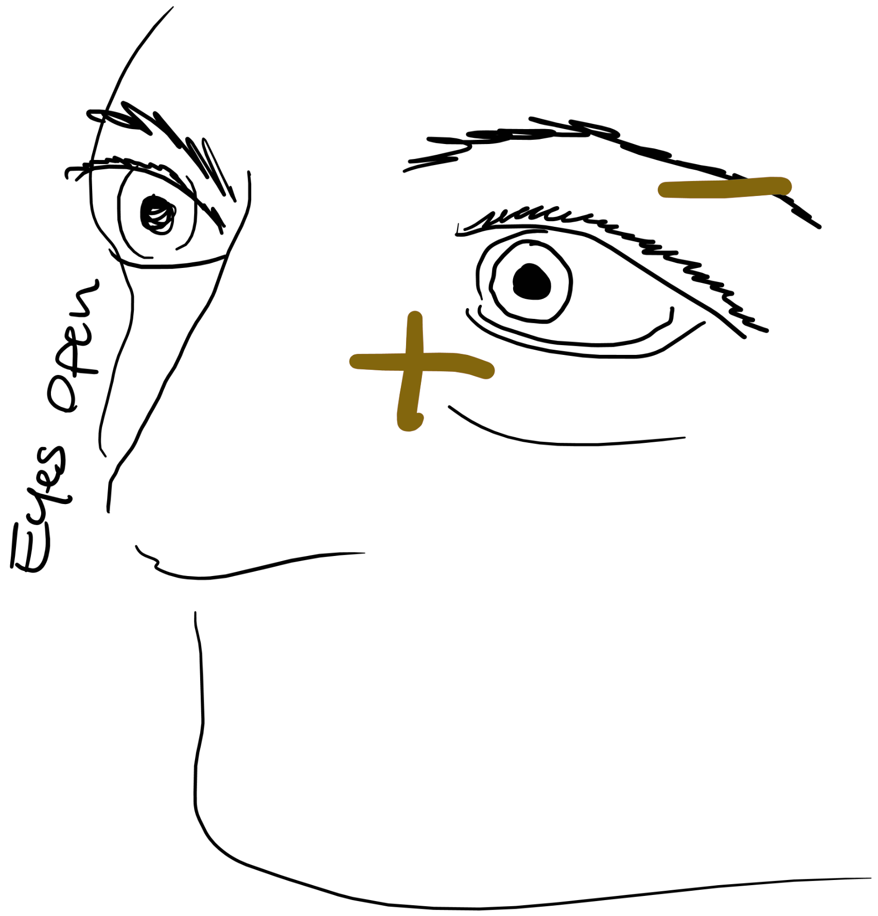
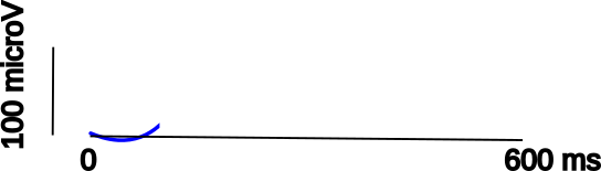 Usual negativity at the back due to the corneoretinal potential
Eye blink as seen in the EOG
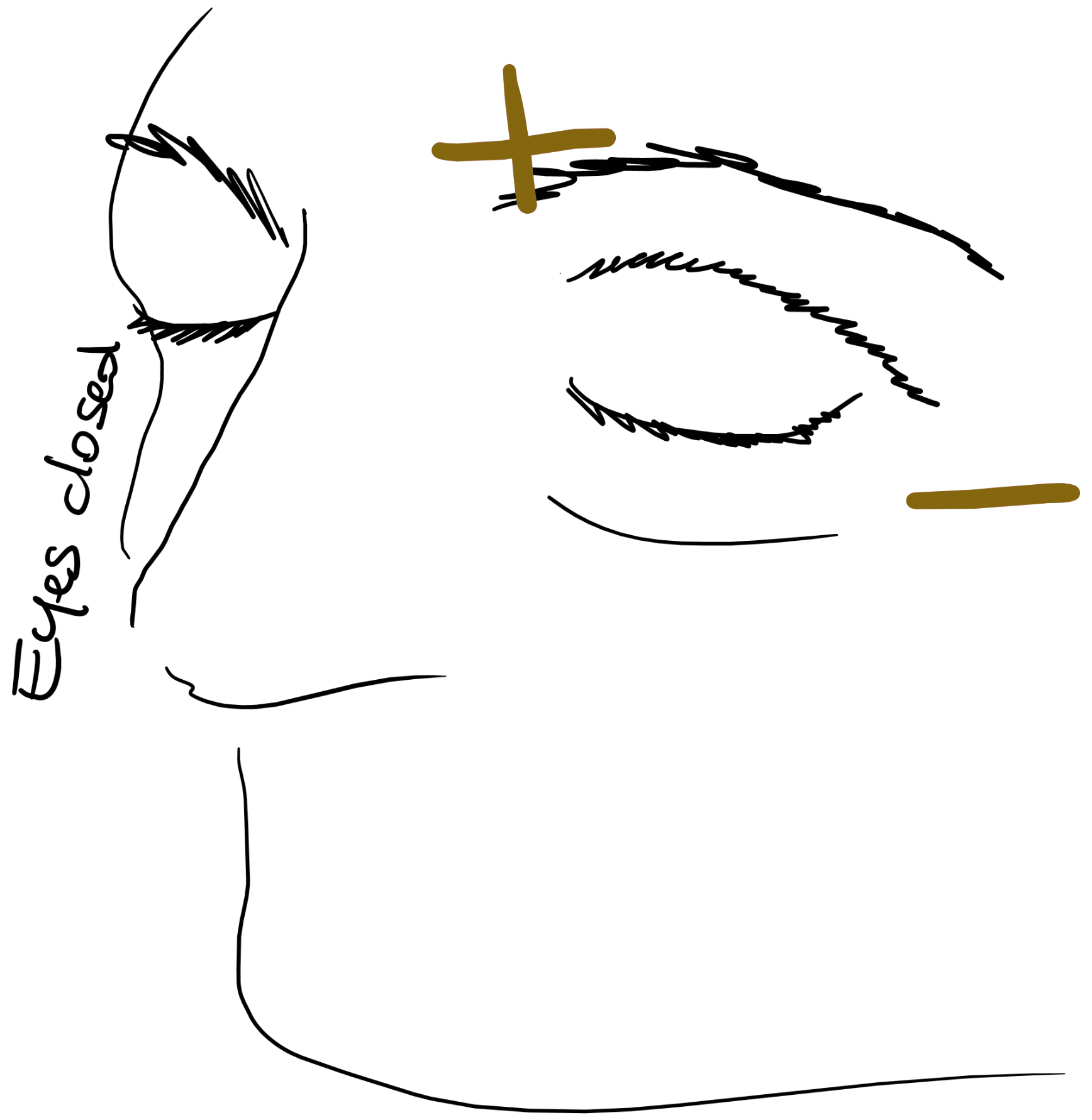
 Transient positivity above the eye
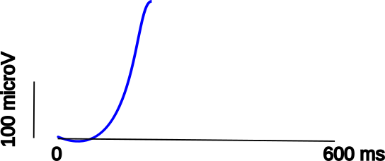
Eye blink as seen in the EOG
 Back to baseline