On a computer press F11 to de/activate full-screen view.
For smartphone and review: Bottom left menu → Tools → PDF Export Mode.
For pdf document: use “learning resources” link above.
Last modified: 2025-11-24
QR code to these slides:
PIN
0000
Agenda
Describe structure and function of:
Neuronal cells (microscopic level)
Nervous system (macroscopic level)
In other words:
How do neurons communicate? Structure and function of neurons
What do neurons communicate? The type of information sent and received
Why do neurons communicate? The higher-level functions that emerge from this communication
Why study the brain, neurons, etc?
A fundamental principle in Biology: “structure determines function”
Psychology studies mental “functions”
Useful for psychologists to understand (at the least at a basic level) the biological substrate to the mental functions.
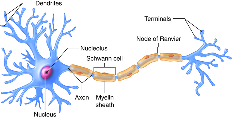
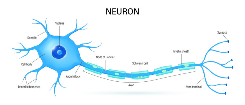
Structure of a neuron
the three main neuronal structures:
soma
dendrites
axon
Drawing of a neuron and its main structures
The soma, or cell body, is the central part of a neuron that contains the nucleus, which houses the cell’s genetic material (DNA). The soma is responsible for maintaining the neuron’s health, regulating its metabolic functions, and synthesizing proteins necessary for the neuron’s structure and signaling processes.
Neurites are projections from the soma and include both dendrites and the axon.
Dendrites are branched extensions that receive signals from other neurons and transmit them toward the soma.
The axon is a long, singular projection that transmits signals away from the soma to other neurons, muscles, or glands.
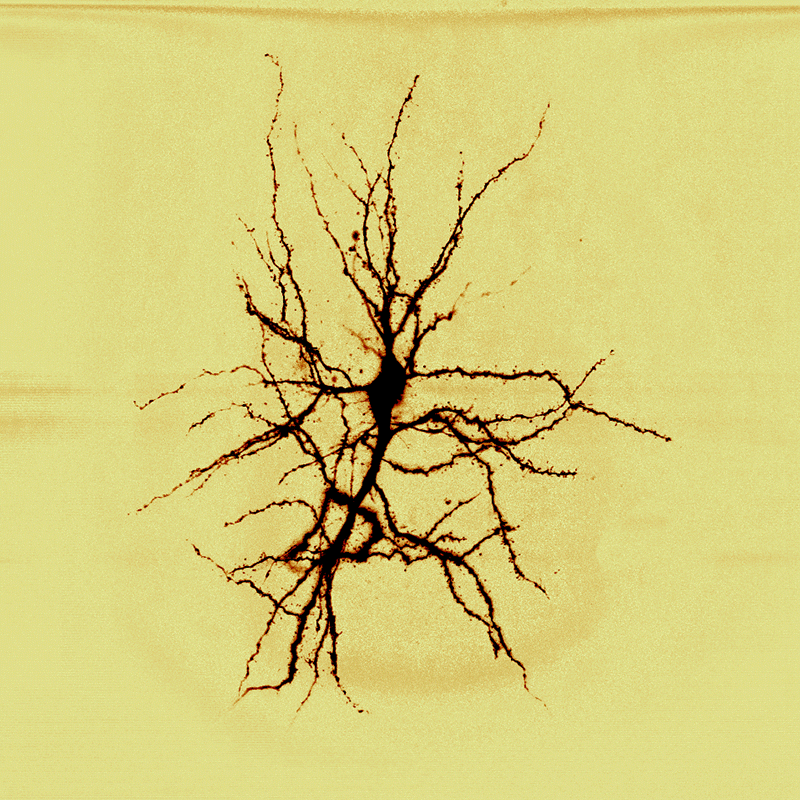
Dendrites
Image of a stellate neuron
Dendrites are specialized structures that receive information* from other neurons (axons).
Each neuron typically has many dendrites, which are organized in a branching pattern known as the dendritic tree.
The structure and shape of dendrites vary across different types of neurons, enabling diverse functional roles in neural communication.
Axon
Electrochemical Communication
The axon is a singular, elongated projection that transmits information away from the soma to other neurons (dendrites or soma).
While each neuron has only one axon, it may branch into collaterals to communicate with multiple targets.
Many axons are wrapped in myelin, a fatty substance that enables faster conduction of electrical impulses by insulating the axon membrane.
Soma
Neuron Cell Body
The soma, also known as the cell body, is the central part of a neuron.It contains the nucleus, which houses the cell’s genetic material,and is responsible for regulating the neuron’s activities, including protein synthesis and energy production.
The soma integrates incoming signals from the dendrites and determines whether to generate an action potential that travels down the axon.
review questions
Which part of the neuron mainly receives information from other neurons?
Axon
Soma
Myelin
Dendrites
Correct response: d
Note the emphasis on mainly in the question: the soma can also receive information but it’s not its main function
When stimulated, the dendrites change their chemicals, generating an electrical post-synaptic potential.
This potential is conducted passively towards the soma and then to the axon hillock.
The potentials generated by many dendrites are summed (some positive, some negative). If the resulting potential is large enough, it triggers an action potential in the axon.
Electrochemical Communication: Action Potential
Image of a neuron with its axon in foreground
The action potential is a rapid change in electrical potential that propagates along the axonand reaches the synapse, the interface (or junction) with another neuron.
Synapse (Pronounced ˈsɪn.æps)
Synapse
The synapse is the junction between the axon terminal of one neuron and the dendrite (or soma) of another neuron.
When an action potential reaches the axon terminal, it triggers the release of neurotransmitters into the synaptic cleft.
These chemicals bind to receptors on the post-synaptic neuron, causing changes in its electrical state.
It is the site where neurotransmitters are released to allow communication between neurons.
Synapses can be excitatory or inhibitory, depending on the type of neurotransmitter and receptor involved. This process is essential for all forms of neural communication, including learning, memory, and reflexes.
The presynaptic neuron (sender) releases neurotransmitters from its axon terminal into the synaptic cleft, whereas the postsynaptic neuron receives and processes the signal.
This interaction can either excite or inhibit the postsynaptic neuron, depending on the type of neurotransmitter and receptor involved.
The summation of multiple post-synaptic potentials will determine whether that neuron will, in turn, generate its own action potential.
Neurotransmitters
Synapse Schematic
Neurotransmitters are molecules that play a crucial role in synaptic communication. They are released by the presynaptic neuron into the synaptic cleft in response to an action potential.
Once released, neurotransmitters bind to specific receptors on the postsynaptic neuron, triggering changes in its electrical or chemical state.
Neurotransmitters: types of functions
Neurotransmittes can either:
excite
inhibit
modulate
the post-synaptic neuron
Excitatory neurotransmitters (e.g., glutamate) increase the likelihood of the postsynaptic neuron firing an action potential.
Inhibitory neurotransmitters (e.g., GABA) decrease the likelihood of the postsynaptic neuron firing.
Modulatory neurotransmitters adjust properties (e.g., excitability) of neural circuits.
The interplay between inhibition and excitation ensures precise and regulated communication among neurons, enabling complex processes such as learning, memory, and motor control.
Review questions
What is the gap between two neurons called?
Explain the difference between pre-synaptic and post-synaptic neurons in synaptic communication.
Types of Neurons
Neurons can be classified based on several characteristics, including:
number of dendrites
shape of dendrites
axon length
type of neurotransmitter they release
This classification highlights the diversity of neurons and their specialized roles in the nervous system.
Classification by Number of Dendrites
Type
Description
Examples
Unipolar
Single projection (one neurite) from the soma that branches into dendrites and an axon.
Sensory neurons
Bipolar
One axon and one dendrite (two neurites) extending from the soma.
Retinal neurons
Multipolar
One axon and multiple dendrites, the most common type of neuron.
Motor neurons, interneurons
Classification by Number of Dendrites
Neuron Shape Classification
Illustration of unipolar, bipolar, or multipolar neurons, depending on the number of projections extending from the soma.
Classification by Shape
Type
Description
Examples
Pyramidal
Triangular-shaped soma with a long apical dendrite and multiple basal dendrites.
Found in the cerebral cortex
Stellate
Star-shaped neurons with radiating dendrites.
Found in the cerebellum
Classification by Shape
Pyramidal and Stellate Cells
Illustration of pyramidal and stellate neurons, highlighting their distinct shapes and structural features.
Classification by Axon Length
Type
Description
Examples
Golgi Type I
Long axons that extend far from the soma.
Motor neurons
Golgi Type II
Short axons that remain close to the soma.
Interneurons
Golgi Type I neurons, also known as projecting neurons, have long axons that extend to distant targets, enabling communication across different regions of the nervous system.
Golgi Type II neurons, also known as local circuit neurons, have short axons that remain close to the soma, facilitating communication within a localized area.
Classification by Neurotransmitter
In addition to excitatory (i.e., they activate the postsynaptic neuron and increase the likelihood of an action potential)and inhibitory (i.e., they suppress the activity of the postsynaptic neuron and reducing the likelihood of an action potential),some neurotransmitters are modulatory: they influence the overall activity of neural circuits by altering the strength or dynamics of synaptic communication.
For example, a modulatory neurotransmitter can influence the excitability of a postsynaptic neuron.
Type
Description
Examples
Excitatory
Release neurotransmitters that increase the likelihood of action potentials in the postsynaptic neuron.
Glutamatergic neurons
Inhibitory
Release neurotransmitters that decrease the likelihood of action potentials in the postsynaptic neuron.
GABAergic neurons
Modulatory
Release neurotransmitters that modulate the activity of other neurons.
Dopaminergic, serotonergic neurons
Dual Nature of Neural Information Communication
Dendrites receives information.
The soma receives and integrates information.
The axon sends information.
But what information are we talking about?
Electrico-chemical potentials
Electrico-chemical potentials
Within a neuron, information is transmitted electrically (post-synaptic potentials, action potential).
Between neurons (in the synapse), information is transmitted chemically via neurotransmitters.
Neurons communicate information using both electrical and chemical mechanisms.
Spiking Rate and Neural Coding
The action potential is a all-or-nothing event (either sent or not sent): same voltage and same duration for each neuron under stable conditions.
So how do neurons communication different information?
Through spiking rate: the number of action potentials sent per second. The frequency at which action potentials are sent (i.e., how often they are sent) can vary.
The spiking rate encodes meaning in neuronal communication.
For example, a neuron may exhibit a high spiking rate during specific activities, such as processing auditory information during speech, indicating its specialization in that domain.
Neurons that respond to similar types of information are often spatially grouped together, reflecting regional functional specialization.
Review questions
Explain the dual nature of neural communication (electrical vs. chemical)
If all action potentials are the same, how do neurons communicate different types or intensities of information?
Not just neurons
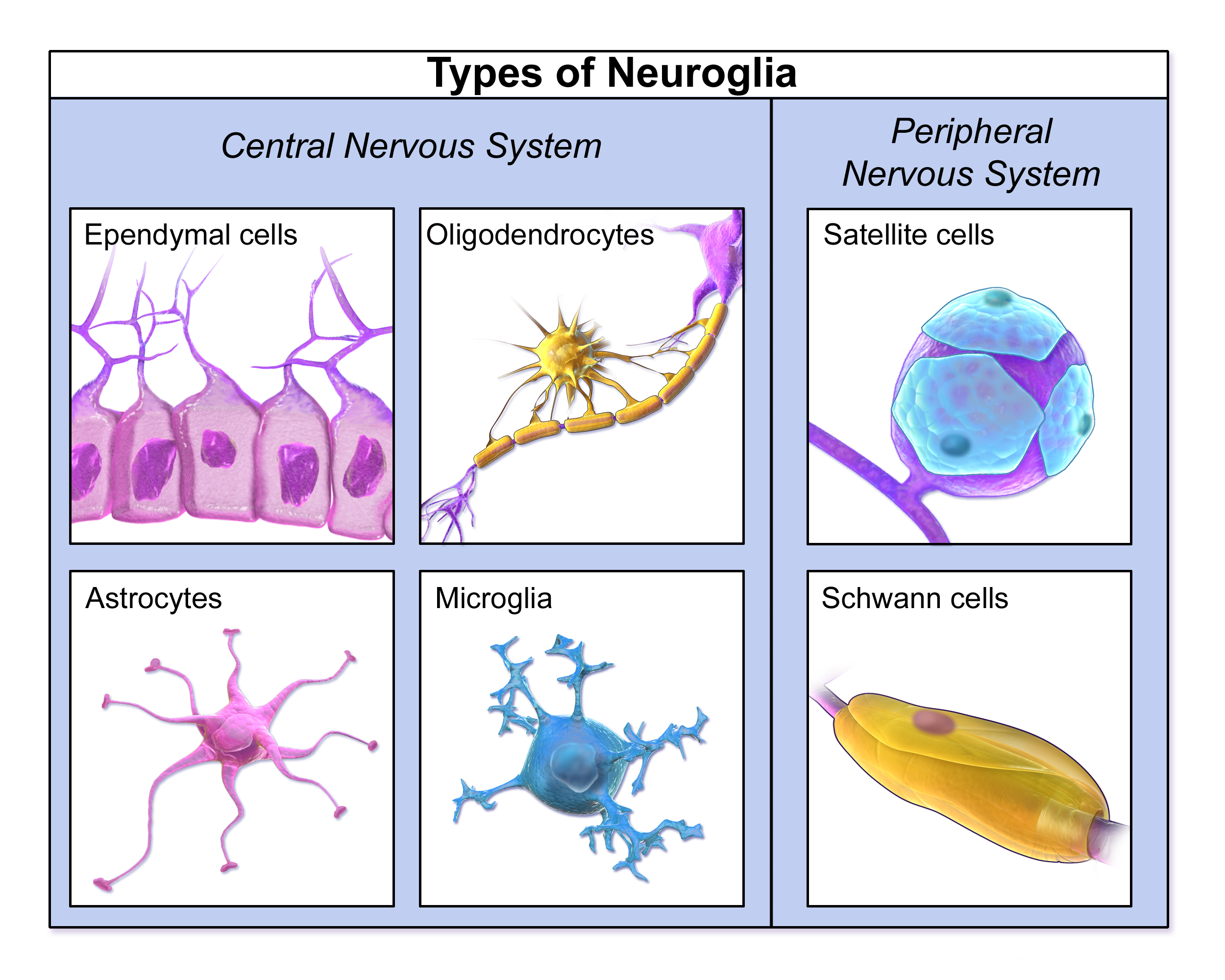
Types of Neuroglia
Glial cells, or glia (pronounced ˈɡliː.ə), are non-neuronal cells in the nervous system that provide support, protection, and nourishment to neurons. They play a critical role in maintaining the homeostasis of the neural environment and facilitating efficient neural communication.
This illustration shows different types of glial cells and their roles.
Types of glia
Brief overview of some glial cells
Type
Function
Astrocytes
Maintain the blood-brain barrier, regulate nutrient supply, and support synaptic function.
Oligodendrocytes
Produce myelin sheaths that insulate axons in the CNS, enabling faster signal transmission.
Schwann Cells
Produce myelin sheaths in the peripheral nervous system (PNS)
Microglia
Act as immune cells in the CNS, clearing damaged cells and protecting against pathogens
Neurons and glia
Until recently, it was believed that the ratio of neurons to glial cells in the human brain was approximately 1:10, meaning there were ten times as many glial cells as neurons. However, recent research has revised this estimate, suggesting a ratio closer to 1:1.
This ratio is not uniform across the nervous system; different regions exhibit varying proportions of neurons and glial cells. For example:
The cerebral cortex has a higher density of neurons relative to glial cells.
The white matter contains more glial cells due to the abundance of myelinated axons.
White and gray matter
Gray and White Matter of the Cerebrum
The brain is composed of gray matter and white matter, each playing distinct roles in neural function.
Gray matter consists primarily of neuronal cell bodies, dendrites, and glial cells. It is involved in processing and integrating information.
White matter is made up of myelinated axons, which facilitate the rapid transmission of signals between different brain regions.
Interim summary
End of the brief “microscopic” view of the brain
Video to watch in your own time, summarizing some of the content covered so far.
Nervous system
The central nervous system includes the brain and the spinal cord.
The cerebrum
The cerebrum includes multiple structures
The cerebral cortex
The outermost layer of the brain, responsible for cognitive functions such as perception, decision-making, language, and conscious thought. It is divided into four lobes:
Cerebral Cortex Lobes
Frontal Lobe: Involved in reasoning, planning, and voluntary movement.
Parietal Lobe: Processes sensory information like touch and spatial awareness.
Temporal Lobe: Handles auditory processing and memory.
Occipital Lobe: Dedicated to visual processing.
Cerebral cortex and cognitive functions
The cerebral cortex enable complex cognitive functions such as:
Perception: processing sensory input from various modalities (e.g., hearing, vision, touch, taste) to create a coherent representation of the environment.
Attention: it allows to select relevant stimuli while filtering out distractions
Memory: encoding, storing, and retrieving ot memories.
Language: speech production and comprehension.
Decision-Making: reasoning, problem-solving, and planning, which are essential for making informed decisions.
Conscious Thought: self-awareness and the ability to reflect on one’s thoughts and actions.
Frontal lobe
The frontal lobe is the largest of the brain’s lobes and is responsible for a wide range of functions, including:
Executive Functions: Planning, decision-making, problem-solving, and goal-setting.
Motor Control: Houses the primary motor cortex, which controls voluntary movements.
Language Production: Includes Broca’s area, essential for speech production.
Attention and Focus: Regulates concentration and the ability to shift focus between tasks.
Emotional Regulation: Plays a role in managing emotions and social behavior.
Working Memory: Supports short-term memory and the manipulation of information.
Personality: Influences traits such as self-awareness, motivation, and social interactions.
Temporal lobe
The temporal lobe is located on the sides of the brain, near the ears, is responsible for functions including:
Auditory Processing: Houses the primary auditory cortex, which processes sounds and enables hearing.
Language Comprehension: Includes Wernicke’s area, essential for understanding spoken and written language.
Memory Formation: Plays a key role in forming and retrieving long-term memories, particularly through the hippocampus.
Emotional Processing: Involved in interpreting emotions and social cues, with contributions from the amygdala.
Object Recognition: Helps identify and categorize objects and faces through visual processing pathways.
Learning: Supports the acquisition of new information and skills.
Parietal lobe
The parietal lobe is located near the top and back of the brain and is involved in various sensory and integrative functions:
Sensory Processing: Interprets sensory information such as touch, temperature, pain, and pressure.
Spatial Awareness: Helps in understanding spatial relationships and navigating the environment.
Proprioception: Processes information about the position and movement of the body.
Integration of Sensory Input: Combines information from different senses to create a coherent perception of the world.
Attention and Focus: Plays a role in directing attention to relevant stimuli.
Mathematical and Logical Reasoning: Supports numerical processing and problem-solving tasks.
Occipital lobe
The occipital lobe is located at the back of the brain and is primarily responsible for visual processing. Its functions include:
Visual Perception: Processes visual information received from the eyes.
Color Recognition: Interprets and distinguishes different colors.
Motion Detection: Identifies and tracks movement within the visual field.
Spatial Processing: Helps in understanding spatial relationships and depth perception.
Object Recognition: Assists in identifying shapes, patterns, and objects.
Visual Memory: Stores and retrieves visual information for future reference.
Review question
An athlete suffers a concussion affecting the frontal lobe. Based on the functions of this lobe, what cognitive or motor difficulties might they experience?
Layers of the cerebral cortex
The cerebral cortex is highly folded, increasing its surface area and enabling complex neural processing.
Structure of the Cerebral Cortex
The cerebral cortex is organized into six distinct layers, each with unique types of neurons and connections.
These layers vary in thickness and composition across different parts (structural specialization), reflecting the functional specialization of each region.
Brodmann areas
Brodmann Areas
The cerebral cortex is divided into distinct zones based on differences in cytoarchitecture. These zones, known as Brodmann areas, identified in the early 20th century by Korbinian Brodmann.
Generally, each area is associated with specific functions, such as sensory processing, motor control, or higher cognitive tasks.
For example:
Primary motor cortex (Brodmann area 4): Contains large pyramidal neurons critical for voluntary movement.
Broca’s area (Brodmann areas 44 and 45): Located in the frontal lobe, it is critical for speech production and language processing.